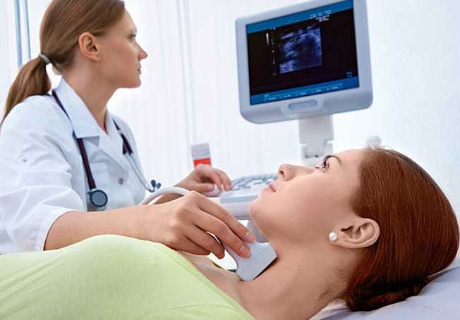
Ultrasound Neck & Thyroid

Modern high-resolution ultrasound has excellent spatial and contrast resolution for the near field, and the development of 3D technology, extended field-of-view or panoramic imaging, and color flow and its diagnostic utility and accuracy.
It is used to see any swelling in the neck like enlarged cervical nodes,enlarged parotid gland,enlarged submandibular gland,any cheek swelling,tumors arising from the muscles,tumors from the blood vessels etc.
read more….
The superficial nature of the neck structures lends itself readily to ultrasound assessment, and ultrasound plays an increasingly important role in head and neck imaging. Ultrasound can provide reliable real-time guidance for fine-needle aspiration cytology (FNAC) or core biopsy, and recognition of its versatility and diagnostic accuracy has led to its routine incorporation in head and neck clinics.
A patient presenting with a neck mass is a common clinical scenario. A meticulous clinical history with physical examination usually provides a reasonable clinical diagnosis. Imaging is necessary for accurate diagnosis and assessment of the extent of a lesion’s involvement prior to treatment.
High-resolution ultrasound is an ideal initial imaging investigation for most neck lumps.1 Cross-sectional modalities serve a supplementary role, offering accurate presurgical anatomic localization, particularly for more deep-seated and locally extensive lesions. The differential diagnosis of a neck mass depends on a patient’s age, the anatomic location of the lesion, and its appearance on ultrasound. Lesions in the head and neck are site-specific with the locations of common lesions and their imaging characteristics.
Ultrasound Thyroid (Thyroid Scan)
It helps to see the thyroid gland within the neck.
It does not use ionizing radiation to visualise the thyroid gland.
It is commonly used to evaluate any lumps/nodules or cancer in the thyroid gland.
This procedure does not need any special preparation.

